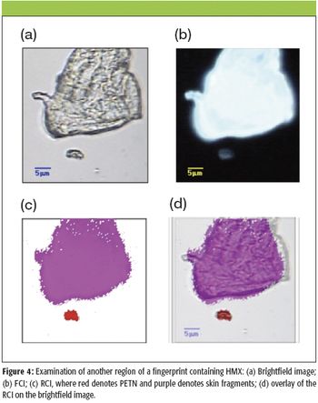
Spectroscopy
The authors discuss the use of Raman spectroscopy to identify an unknown gem or mineral sample or to verify that a known sample has been classified correctly.


Spectroscopy
The authors discuss the use of Raman spectroscopy to identify an unknown gem or mineral sample or to verify that a known sample has been classified correctly.

Special Issues
This article discusses instruments that can be used in the field to rapidly and accurately identify various explosives and their precursors.

Spectroscopy
In this article, columnist Fran Adar will review the important features of the Raman spectra of these materials and indicate why the extracted information is important for material development and engineering.

Spectroscopy
Columnist Fran Adar discusses the physical determinants of spatial resolution and developing methods to improve the mapping speeds of Raman images.

Special Issues
The authors discuss the use of instruments that combine FT-IR and Raman microscopy on a single platform, enabling synergistic studies of many materials.

Spectroscopy
Surface-enhanced Raman spectroscopy (SERS) has experienced an explosive resurgence in interest lately. Development of reproducible, spatially uniform SERS-active substrates has made this technique an attractive approach for identification of Raman-active compounds and biological materials including toxins, intact viruses, and intact bacterial cells–spores...

Special Issues
Raman spectroscopy is going through a major revolution with the continuous introduction of new fiber-based modular systems for low-resolution applications. More and more scientists are discovering what Raman spectroscopy can do for their research, education, and commercial applications thanks to the low costs and flexibility this new technology is providing. New applications and prospects are presented each day, and it is important to understand the advantages and limitations that this user-friendly analytical technique can provide to address these opportunities with a scientific approach.

Special Issues
Over the last few years, Raman has made the transition from a technique used solely in a research environment to one that is now seen as a powerful tool for routine analytical use. Raman spectroscopy now is used widely for sample identification in fields as diverse as forensics, QA/QC, art conservation, defect analysis, and failure analysis. This has imposed new demands on the technique for reproducibility and stability. Successful sample identification takes advantage of the extensive spectral libraries and sophisticated search algorithms that have been developed in recent years. However, in order to be able to cross-correlate experimental and library spectra with any degree of confidence, it is critical that the Raman spectrometers used to collect the spectra are calibrated rigorously. It is likewise critical for QC applications that spectra collected on one instrument can be compared reliably with spectra collected on other instruments and that results remain constant when collected over extended periods..

Special Issues
Raman imaging has moved on. It is now possible to capitalize on the wealth of information available from a Raman spectrum by imaging materials over large areas, with the spatial resolution, spectral resolution, and laser excitation parameters tailored to suit each application. Raman experiments and images from a diverse range of samples are presented.

Special Issues
Chemical analysis in the forensic field is different in many aspects from other areas of analysis. The ultimate goal is to identify the sources of evidence, often by matching chemical composition. In this regard, identifying minor elements or trace impurities is as important as identifying main ingredients. In some cases, identifying minor and trace components can be critical to determining that material collected at the site of a crime is identical to material collected in a suspect's environment. In other cases, full identification of trace evidence can be important. Raman microscopy is capable of providing both types of information on minute amounts of material.

Special Issues
Since it was first described in 1974, surface-enhanced Raman spectrometry (SERS) has been thought to offer significant potential for a range of different applications. The theoretical sensitivity and specificity envisaged for this powerful technique has engaged scientists for many years, but practical challenges have hindered its routine adoption. Now, a new approach combines a robust and reliable substrate with expertise in surface chemistry and molecular biology on a platform that can be adapted for a wide variety of Raman instrumentation and customized routine applications.

Special Issues
The combination of confocal Raman and atomic force microscopes allows chemical and surface topography imaging of large samples without any ongoing process control by an operator. This article describes the relevant measurement principles and presents examples of automated measurements on nanostructured materials.

Spectroscopy
The continuing pace of technological advancements in scientific instruments has recently led to a wide range of commercially viable portable and handheld instruments, and the Raman spectroscopy market is no exception. While security applications have received much of the early attention in relation to handheld instruments, other applications are beginning to replace demand from the security markets.


Spectroscopy
Chemical analysts who use spectroscopy to extract molecular information from samples have been following the developments in Raman instrumentation. Vibrational spectroscopy provides detailed molecular information, but Fourier-transform IR has been much easier to use than Raman. Now that Raman equipment is smaller, cheaper, faster, and easier, analysts are interested. Columnist Fran Adar will discuss why.

Application Notebook

Spectroscopy
A new method for the early detection of gastric cancer uses a combination of feature extraction based upon continuous wavelets for Fourier transform-infrared spectroscopy (FT-IR) and classification using an artificial neural network trained with a back-propagation algorithm.

Spectroscopy
Raman microscopy was developed as a tool for microanalysis complementary to the electron microscope, which enabled identification of the elements in a microspot. The first realization for Raman imaging was implemented using a nonconfocal optical method. Subsequently, a confocal scheme was developed, which provided better contrast in the Raman image. A number of successful examples from pathology, pharmaceutical analysis, and geology will be shown.

Spectroscopy
Raman microspectroscopy is a powerful tool for noninvasive chemical analysis of tissues, cells, and cellular structures. To achieve the highest signal-to-noise ratio and fidelity of Raman spectra, the background must be minimized. The difference in temporal dependence of Raman and fluorescence signals can be used for very effective discrimination. A careful system design, based upon the employment of very efficient Kerr-gating materials, makes confocal Raman microscopy possible with significantly shorter acquisition times. The new instrument is tested for a variety of biomedical systems. The possible applications are outlined together with the routes for further improvement.

Spectroscopy
Columnist Fran Adar discusses the evolution of Raman spectroscopy instrumentation and new applications for this more sensitive, easy-to-use technology.

Application Notebook
Lambda Solutions, Inc.


Spectroscopy
In this investigation the authors assess the potential of Raman spectroscopy as a tool for probing conformational changes in membrane-spanning proteins - in this case, the sodium potassium adenosine triphosphatase (Na+,K+-ATPase).

Spectroscopy
November 2006. Raman spectroscopy is a promising new tool for noninvasive, real-time diagnosis of tissue abnormalities. Here, we show evidence of its application for cancer diagnosis in four distinct tissue types: skin, breast, gastrointestinal tract, and cervix. Multivariate statistical analysis and discrimination algorithms allow for automated classification of the spectra into clinically relevant pathological categories using histology as a gold standard. Although limitations exist, the technique shows every indication of being an exciting prospect in the management of cancer in a clinical setting.

Spectroscopy
September 2006. The authors rapidly acquire complete vibrational spectra in the fingerprint region using a single femtosecond laser for broadband coherent anti-Stokes Raman scattering (CARS) microscopy to image spatially variant compositions of condensed-phase samples.