

An interview with 2014 Charles Mann Award winner Richard P. Van Duyne, the Charles E. and Emma H. Morrison Professor of Chemistry at Northwestern University in Evanston, Illinois.
Raman Spectroscopy
Latest News
Advertisement
Advertisement
Dustin McIntyre, of the National Energy Technology Laboratory, US Department of Energy in Morgantown, West Virginia, has been exploring the use of laser-induced breakdown spectroscopy (LIBS) to measure subsurface gases, liquids, and solids at subsurface conditions.

Spectroscopy
Polarized Raman spectra are presented along with a discussion of the association of the symmetry species of the normal vibrational mode and the depolarization ratio of Raman scattering.
Spectroscopy
Spectroscopy magazine is seeking contributed manuscripts for its June 2014 supplement on Raman spectroscopy.
Everyone loves a list, and the editors of Spectroscopy are no exception! In 2013, Spectroscopy covered a wide array of topics throughout the year to bring you the most relevant information for your work, on topics ranging from selecting the right ICP-MS system to deciding which Raman technique is right for you, from our annual salary survey to calibration transfer. Here is a list of 13 popular articles and columns from 2013
In this interview with Spectroscopy, renowned planetary spectroscopist Alian Wang of Washington University in St. Louis discusses the development of the Mars microbeam Raman spectrometer (MMRS), proposed to be part of science payload for the NASA-funded mission, Mars 2020. Wang discusses the challenges involved in making the instrument robust enough for operation in space, and highlights how data collected from over 18 years has advanced our understanding of the history of Mars.
Click here to view the complete Wavelength newsletter from December 17, 2013.
In this interview with Spectroscopy, renowned planetary spectroscopist Alian Wang of Washington University in St. Louis discusses the development of the Mars microbeam Raman spectrometer (MMRS), proposed to be part of science payload for the NASA-funded mission, Mars 2020. Wang discusses the challenges involved in making the instrument robust enough for operation in space, and highlights how data collected from over 18 years has advanced our understanding of the history of Mars.
Spectroscopy
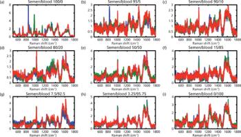
In this study, regression and classification chemometrical algorithms were combined to achieve effective discrimination of pure body fluids from their binary mixtures.
Spectroscopy
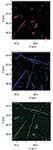
A summary of the efforts to study microorganism biofilms using surface-enhanced Raman scattering, a powerful technique for in situ analysis of biological molecules and biomolecular organizations
Spectroscopy
The physics that determine how gratings and spectrographs work are summarized in simple terms for new users of Raman equipment.
Click here to view the complete Wavelength newsletter from September 10, 2013.

An interview with Paul Pudney, a winner of the William F. Meggers Award.
Spectroscopy
What exactly is a "Raman image" and how is it rendered? The authors explain those points, and demonstrate the use of Raman imaging for the characterization of thin-film and ion-implanted silicon structures. High spectral resolution makes it possible to resolve or contrast the substrate silicon and polysilicon film in Raman images and thus aids in the chemical or physical differentiation of spectrally similar materials.
Click here to view the complete Wavelength newsletter from June 19, 2013.
Spectroscopy
The presence of electronic transitions in the visible part of the spectrum can provide enormous enhancement of Raman signals.
Special Issues
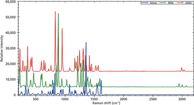
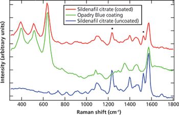
A modern portable Raman instrument equipped with a 1064-nm laser is compared to one using a 785-nm laser for the identification of counterfeit pharmaceuticals.
Special Issues
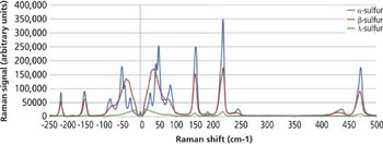
Results are reported from studies using volume holographic grating filter technology with both visible and NIR excitation wavelengths to analyze multiple forms of the polymorphic active pharmaceutical ingredient carbamazepine.

Special Issues
This article re-explains and demystifies some definitions and opinions concerning Raman spectroscopy from two distinct sides: academic and industrial.
Special Issues
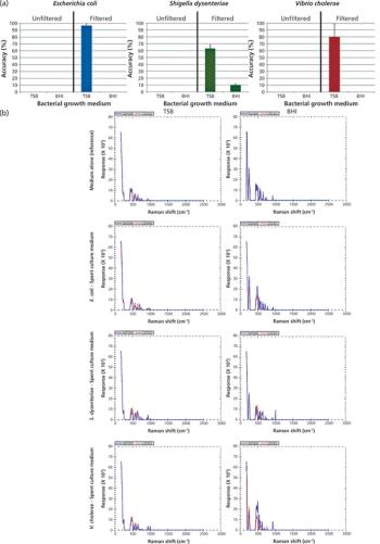
The feasibility of identifying different types of bacterial growth media using a handheld Raman instrument with a proprietary baseline correction algorithm is investigated.

Spectroscopy
Volker Deckert, the winner of the 2013 Charles Mann Award, is advancing the use of tip enhanced Raman spectroscopy (TERS) to push the lateral resolution of vibrational spectroscopy well below the Abbe limit, to achieve single-molecule sensitivity. Because the tip can be moved with sub-nanometer precision, structural information with unmatched spatial resolution can be achieved without the need of specific labels.

With improvements in instrumentation, Raman spectroscopy continues to expand its range of applications to diverse areas of materials analysis and research.
Spectroscopy
Here, real spectra illustrate how to be your own critic when evaluating band-fitted spectra.
Spectroscopy
Micro-Raman spectroscopy has been used to depth-profile a waveguide produced by an ion-exchange reaction in a single crystal of a ferroelectric metal oxide, and to reveal the changes in chemical bonding and atomic structure that occur in this process.
Special Issues
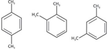
This tutorial demonstrates that Raman is well-suited to tackle two challenges faced by industry: complex mixtures and isomers.
Advertisement
Advertisement
Trending on Spectroscopy Online
1
The State of Spectroscopy Careers
2
Exploring Mars' Lost-Water Narrative Using Raman Spectroscopy and X-ray Fluorescence
3
From Single-Technique Analysis to Multimodal Characterization: Recent Advances and Future Perspectives for Carbon Nanotubes and Graphene
4
Is Produced Water a Source of Lithium?
5