

Spectroscopy Interviews
Latest News
September 13th 2019
By John Chasse
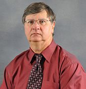
Spectroscopy can be difficult to carry out outside a controlled laboratory environment. Imagine, then, the hurdles that would accompany performing spectroscopy in the extreme conditions of deep space or the ocean floor. Mike Angel, a professor of chemistry at the University of South Carolina, has taken on those challenges, working on new types of instruments for remote and in- situ laser spectroscopy, with a focus on deep-ocean, planetary, and homeland security applications of deep ultraviolet Raman, and laser-induced breakdown spectroscopy to develop the tools necessary to work within these extreme environments.
Advertisement
Latest Videos
Shorts
1:20
How to Stop Microplastics: From Washing Machines to Storm Drains
8 months ago
by
Will Wetzel
0:38
The Treatment of Water Leads to Microplastics in Bottled Water
8 months ago
by
Will Wetzel
1:00
Capturing Microplastics off Car Tires
8 months ago
by
Will Wetzel
1:23
The Dirty Secret of 'Dirt Cheap' Lithium Sulfur Batteries
9 months ago
by
Johanna Nelson Weker
0:41
We Separate Cancer Cells By Physics, Not Antibodies
9 months ago
by
Will Wetzel(+1 more)
0:46
Universal Sensors Are Like Radios Without Tuning Knobs!
9 months ago
by
Steven Bell
0:50
The Key to Selective Actinide Extraction
9 months ago
by
Robert Lascola
1:19
ML & Spectroscopy: The Future of Pathology
9 months ago
by
Rohith Reddy
0:52
Seeing The Raman Signal From 1-3 Molecules
9 months ago
by
Zachary D. Schultz
0:40
Not Your Grandfather’s NMR
9 months ago
by
Damodaran Krishnan Achary
Advertisement
More News
Timothy J. Johnson and Tanya L. Myers, the 2019 winners of the Applied Spectroscopy William F. Meggers Award, have been exploring novel methods using infrared (IR) and visible reflectance spectroscopies for identification of target chemicals through derivation of the n and k optical constants. For their winning paper, Johnson and Myers created a spectroscopic library of 57 liquids for which they measured the complex refractive index, data that can be used for optical modeling and other purposes. They shared some of the details of their work with Spectroscopy.
Barry Wise, the president of Eigenvector Research, is a renowned expert in chemometrics. We recently spoke to him to get his thoughts and opinions on the latest developments in chemometrics for the analytical sciences.
Spectroscopy
SciX Award winner Benedikt Schwarz explains his groundbreaking work in portable infrared devices.
Spectroscopy
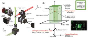
Javier Laserna has developed a technique for analyzing the composition of individual nanoparticles that is capable of detecting nanoparticles as the attogram level, by combining LIBS with optical trapping.
Raman spectroscopy is a versatile tool to identify and characterize the chemical and physical properties of graphene-based materials (1-4). Raman spectroscopy provides information on graphene structures for fundamental research and for practical device fabrication. Raman scattering demonstrates the first- and second-order modes in intrinsic graphene as well as the shear, layer-breathing, and the G and 2D modes of multilayer graphene. Professor Ping-Heng Tan from the State Key Laboratory of Superlattices and Microstructures at the Institute of Semiconductors at the Chinese Academy of Sciences is carrying out new research to advance the use of Raman analysis of these materials. We recently interviewed Tan about this work.
Spectroscopy
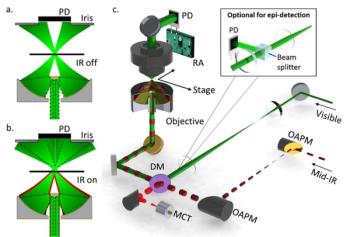
Significant progress is being made to harness the power of spectroscopy technique for medical research. An ongoing challenge, and area of development, in this effort, is to “see” more and more detail about biological activity, even within individual cells. Ji-Xin Cheng, a professor of biomedical engineering at Boston University, is advancing such work, by developing techniques like midinfrared photothermal (MIP) imaging and Raman spectromicroscopy. Cheng is the 2019 winner of the Ellis R. Lippincott Award, which is awarded annually by the Optical Society, the Coblentz Society, and the Society for Applied Spectroscopy, to an individual who has made significant contributions to the field of vibrational spectroscopy. Here, Cheng speaks to us about those techniques.
Recent advances in Raman spectroscopy, specifically using surface enhanced spatially offset resonance Raman spectroscopy (SESORRS), which is a combination of surface enhanced Raman scattering (SERS), and spatially offset Raman spectroscopy (SORS) are enabling noninvasive, real-time measurements of living tissue and multiple bacterial pathogens. In an interview with Karen Faulds, the 2019 recipient of the FACSS Charles Mann Award for Applied Raman Spectroscopy, we explore the latest developments in Raman spectroscopy for biomedical analysis applications. This interview is part of a series of interviews with the winners of awards presented at the SciX conference.
The 2019 recipient of the Clara Craver award, Xiaoyun (Shawn) Chen, is a senior research scientist working in the Core R&D Analytical Sciences department of the Dow Chemical Company. Chen, who will receive this award this fall at SciX 2019 in Palm Springs, California, recently spoke to Spectroscopy about his work.
Spectroscopy
Nick Stone of the University of Exeter explains why mid-infrared (mid-IR) spectroscopy is so valuable for spectral imaging in disease research and clinical diagnostics, and discusses his own recent work in this area.
Spectroscopy
Mohamad Sabsabi of the National Research Council of Canada explains the fundamentals of LIBS, its recent evolution, and its benefits and limitations, and offers advice for new users.
Advanced techniques in tissue engineering hold promise to those who suffer from damage to or degeneration of joint cartilage. But some challenges exist for tissue engineers to gain a better understanding of the development of these constructs and their mechanical properties. Nancy Pleshko, a professor of Bioengineering at Temple University (Philadelphia, Pennsylvania) has been studying the use of Fourier-transform–infrared imaging spectroscopy (FT-IRIS) as well as near-infrared (NIR) spectroscopy to explore the ways in which these techniques can aid in the development of replacement tissue. We spoke to her about her research and findings.
Spectroscopy
Laser-induced breakdown spectroscopy (LIBS) has seen significant expanded adoption in recent years, particularly in industrial applications where it can provide important advantages over other techniques. Mohamad Sabsabi, of the National Research Council of Canada, has been leading programs to research and implement the use of LIBS in mining and energy applications.

Lili He, an associate professor in the Department of Food Science at the University of Massachusetts, Amherst, focuses on developing and applying advanced analytical techniques to solve critical and emerging issues in food science. Recently, that focus has turned to using surface-enhanced Raman spectroscopy (SERS) in the detection, analysis, and identification of both natural and artificial food colorants. Dr. He recently spoke to Spectroscopy about that work.
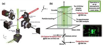
As the use of nanoparticles in a wide range of applications continues to increase, the need for techniques to analyze these particles also increases. Javier Laserna of the University of Málaga, in Spain, has developed an approach to measure individual nanoparticles in air using optical trapping (OT) combined with laser-induced breakdown spectroscopy (LIBS). Laserna received the 2018 Lester W. Strock award for this development as well as his broader research on analytical methods and instrumentation using LIBS. He recently spoke to us about this work.
We recently interviewed Benedikt Schwarz, Ph.D., a principal investigator at TU Wien, in Vienna, Austria, about his work in the integration of a bi-functional quantum cascade laser with detection capabilities for lab-on-a-chip mid-IR absorption spectroscopy. Schwarz won a FACSS Innovation award at the 2018 SciX conference for this work, and this interview is part of a series of interviews with the winners of awards presented at SciX.
Spectroscopy
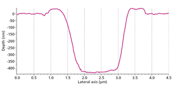
Jorge Pisonero of the University of Oviedo studies the performance of the laser ablation process in laser ablation–inductively coupled plasma-mass spectrometry, examining aspects such as the morphology of the craters created during sampling and the role of laser fluence. He also studies the effect of these factors in specific applications of LA-ICP-MS. He recently spoke to us about this work.
The challenge of tackling neurogenerative diseases like Parkinson’s and Alzheimer’s leads researchers to study the role of metals, for which atomic spectroscopy tools have become essential. Dominic Hare, the 2019 winner of the Emerging Leader in Atomic Spectroscopy award, presented by Spectroscopy, is a forerunner in the use of laser ablation-inductively coupled plasma-mass spectrometry (LA-ICP-MS) to image metals in biological tissue as part of the work to improve our fundamental understanding of neurodegenerative diseases. He recently spoke to us about this work. The award will be presented to Hare at The European Winter Conference on Plasma Spectrochemistry (EWCPS), in Pau, France, February 3-8, 2019.
Spectroscopy
Raman spectroscopy is promising some dramatic breakthroughs in biomedical applications. Juergen Popp and his team are determined to realize that promise, by working to make the technique a powerful tool for cell biology and clinical studies.
Spectroscopy
The 2018 Award winner discusses her career challenges and her research on the use of specially engineered proteins, combined with 2D IR spectroscopy, for investigating protein function dynamics.
On-capillary surface-enhanced Raman spectroscopy (SERS) is showing dramatic potential for analysis of human whole blood constituents using microsampling. A group of researchers has recently published a method to measure glutathione (GSH) in a 2 μL sample of human whole blood. This exciting development could lead to rapid point-of-care analysis of other essential blood components. We recently interviewed Julia Kuligowski of the Health Research Institute La Fe, in Valencia, Spain, and Guillermo Quintas, of the LEITAT Technological Center in Barcelona, about this research.
Megan Thielges, an associate professor of chemistry at Indiana University is the recipient of the 2018 Emerging Leader in Molecular Spectroscopy Award, presented by Spectroscopy magazine. This award, presented at the SciX conference each year, recognizes a uniquely talented young molecular spectroscopist. This October, Prof. Thielges will give a plenary lecture and be honored within a SciX award symposium. She recently spoke with us regarding her research work, and a few other topics, demonstrating the application of site- specific 2D IR spectroscopy for investigating protein function dynamics.
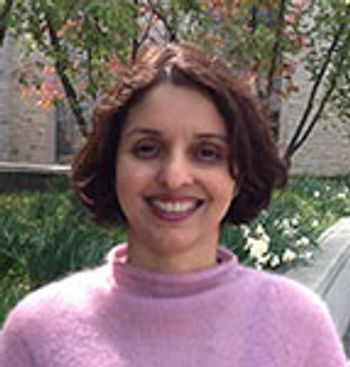
Surface-enhanced Raman spectroscopy (SERS) and surface-enhanced spatially offset Raman spectroscopy (SESORS) have been used in medical research for the detection of neurotransmitters such as melatonin, serotonin, and epinephrine. These techniques can assist in the diagnosis of neurological diseases and provide information that can lead to more effective treatment methods. Bhavya Sharma an assistant professor in the department of chemistry at the University of Tennessee (Knoxville, Tennessee), has been using SERS and SESORS to detect neurotransmitters and probe subsurface layers through the skull. Here, she describes the advantages of these techniques and how they are used in biological applications.
Spectroscopy
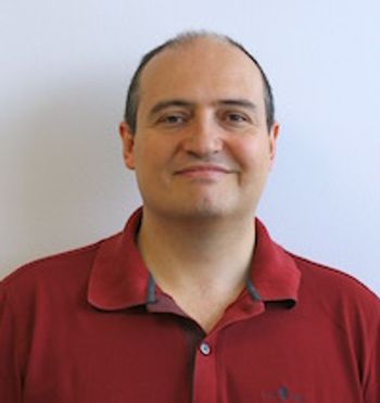
The detection, quantitation, and characterization of nanoparticles using inductively coupled plasma–mass spectrometry (ICP-MS), and in particular using single-particle ICP-MS (SP-ICP-MS), has developed significantly in recent years. However, the difficulties involved in this type of analysis vary, depending on the composition of the nanoparticles. Martín Resano of the University of Zaragoza, together with colleagues from Ghent University, has recently developed a method for characterizing nanoparticles made from silicon dioxide (Si02), which are much more challenging to detect than those made from silver or gold. He recently spoke to us about this work.
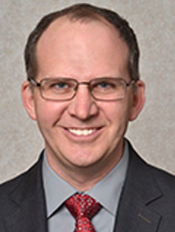
If a new drug candidate is going to fail, it’s best if it does so as early in the process as possible-before a lot of time and money have been spent developing it. Figuring out whether a drug will fail, and why it might fail, is a complex problem, however. . Zachary Schultz of The Ohio State University is investigating how tip-enhanced Raman spectroscopy (TERS) can help with this process, particularly in terms of studying binding between membrane receptors and ligands.
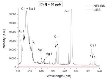
Laser-induced plasmas are formed by the application of a laser pulse to a target surface, which instantly excites, ionizes, and vaporizes the material into a very hot vapor plume. One of the main uses of these plasmas is in laser-induced breakdown spectroscopy, a rapidly evolving and exciting field of study. Alessandro De Giacomo is a professor in the Department of Chemistry at the University of Bari in Italy and an associated researcher at CNR-NANOTEC, and he and his group are involved with the study of laser-induced plasmas and the use of nanoparticles (NPs) in laser-induced breakdown spectroscopy to enhance signal. We recently spoke with him about this research.
Advertisement
Advertisement
Trending on Spectroscopy Online
1
Peter R. Griffiths (1942–2026): A Life Devoted to Infrared and Vibrational Spectroscopy
2
How is Raman Spectroscopy Being Used in Life Science Applications?
3
The Current Gaps in Food Science
4
In Memoriam: John Philip Walters (July 4, 1938 - August 18, 2026)
5