Spectroscopy Interviews
Latest News
Advertisement
Latest Videos


Shorts
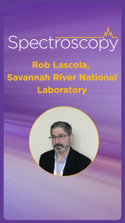
0:57
Controlling Selectivity via Molecular Structure
9 months ago
by
Robert Lascola
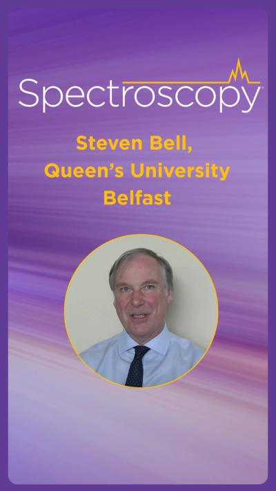
0:57
Why Internal Standards Are Crucial for Accurate Detection
9 months ago
by
Steven Bell
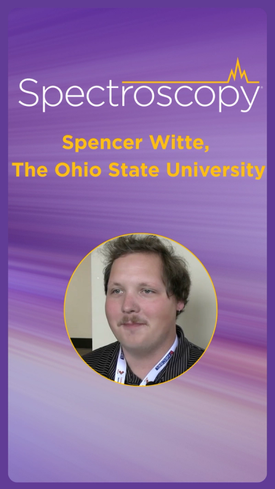
0:41
How Hair Can Reveal Chemical Changes Over Time Using Raman Spectroscopy
10 months ago
by
Spencer Witte
1:04
Revolutionizing Plant Analysis with AI and Spectroscopy
10 months ago
by
Renee Romano
0:42
Watching Chemical Bonds React Inside Living Cells
10 months ago
by
Zachary D. Schultz
0:42
Fusing Spectra to Fix Measurement Errors
10 months ago
by
Robert Lascola
0:48
Why Attending the SciX Conferences Matters
10 months ago
by
Zachary D. Schultz
1:03
Why Do Molecules Naturally Stick to Surfaces?
10 months ago
by
Steven Bell
0:55
How Colored Actinides Ruin Your Raman Spectroscopy Readings
10 months ago
by
Robert Lascola

0:39
The Types of Litter in Lake Tahoe
a year ago
by
Monica Arienzo
Advertisement
More News

Although inductively coupled plasma-optical emission spectrometry (ICP-OES) and ICP-mass spectrometry (MS) are generally considered to be mature techniques, researchers continue to investigate the fundamentals of the techniques and improve their capabilities. Diane Beauchemin, a professor at Queens University in Kingston, Ontario, is engaged in that challenge. She recently spoke to Spectroscopy about methods she has developed for simultaneous speciation and her work to improve sample introduction efficiency, to improve sensitivity and detection limits.
In biology, the study of intracellular structures is important and requires analytical techniques with submicrometer resolution. Atomic force microscopy-infrared (AFM-IR) spectroscopy is one technique that has the required lateral spatial resolution to observe such structures. David Perez-Guaita, PhD, at the Centre for Biospectroscopy at Monash University in Australia, is pioneering work applying AFM-IR to the study of red blood cells infected with the malaria parasite.
Lasers are used for a wide range of industrial, medical, spectroscopic, and military applications. Daniel Kazal, a graduate research assistant in the Department of Chemistry and Biochemistry at the University of Maryland Baltimore County, has developed a novel technique for channeling sound using a tube-shaped laser beam that forms a thermal gradient. Based on his work with this approach, he received the 2017 FACSS Innovation Award. We recently spoke with him about this research. This interview is part of a series of interviews with the winners of awards presented at SciX.

Geochronology is an exciting area of atomic spectroscopy and earth science research. One of the goals is to answer tectonic questions, and in particular, how the crust responds to continent–continent collision. John M. Cottle, a professor of earth science at the University of California, Santa Barbara, is one of the scientists on that mission. Cottle and his research group are at the forefront of discovery in geochronology, combining both laboratory and field-based research. In particular, Cottle is a leader in the development of novel laser-ablation inductively coupled plasma–mass spectrometry (LA-ICP-MS) measurements and their application to tectonic questions in convergent orogens, which are mountain ranges formed when a continental plate crumples and is pushed upwards.
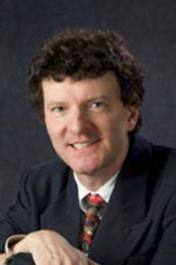
Near-infrared (NIR) spectroscopy is an important technique in the pharmaceutical industry because of its ability to provide information about bulk material without sample preparation. Multivariate calibration techniques are frequently used to analyze the NIR data. Robert Lodder, who is a professor in the Department of Pharmaceutical Sciences at the University of Kentucky in Lexington, Kentucky, uses NIR spectroscopy along with an interesting alternative calibration technique, molecular factor computing, in his work with an experimental drug for combating the Ebola virus. We recently spoke with him about his research.
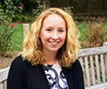
The atomic spectroscopy techniques of laser-induced breakdown spectroscopy (LIBS) and X-ray fluorescence spectroscopy have different strengths. Lydia Breckenridge, a senior research investigator at Bristol-Myers Squibb, uses both techniques in her work in pharmaceutical development. Here, she shares some of the advantages and challenges of using these techniques, and how the greatest benefits are sometimes derived by focusing on their complementarity, and using them in combination.

Two-dimensional (2D) Raman correlation spectroscopy is a powerful analytical technique for analyzing a system under the influence of an external perturbation. Isao Noda, of the Department of Materials Science and Engineering, at the University of Delaware and Danimer Scientific, has been developing 2D Raman correlation spectroscopy and applying it to the study of various materials, including exciting new biopolymers. He recently spoke to us about this work.
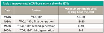
In recent years, researchers have been making important developments to advance the effectiveness of spectroscopic techniques for biomedical uses ranging from the identification of infectious agents to measuring the edges of cancerous tumors. X-ray fluorescence (XRF) spectroscopy is among the techniques that can have useful medical applications. David R. Chettle, a professor in the Department of Physics and Astronomy at McMaster University in Hamilton, Ontario, Canada, uses XRF for the in vivo measurement of toxic elements in human subjects, with the goal of developing devices that can be used to investigate the possible health effects of toxin exposure. He recently spoke to us about his research.

Bioanalysis, and particularly medical diagnostics, is an exciting area of spectroscopy research. One of the dreams is to develop spectroscopic tools that can be used for point-of-care diagnostics with a smartphone. Russ Algar, an assistant professor in chemistry at the University of British Columbia (UBC), in Vancouver, Canada, is one of the scientists on that quest. Algar and his research group focus on the development of nontraditional fluorescent materials-such as quantum dots, luminescent lanthanide complexes, and semiconducting polymer dots-for biochemical sensing. They are studying how these materials can be applied to a variety of problems, including molecular medicine, personalized medicine, and yes, point-of-care diagnostics with smartphones. For his work, Algar has been chosen as the winner of second annual Emerging Leader in Molecular Spectroscopy award, presented by Spectroscopy magazine.
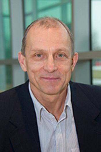
In recent years, there have been significant advances in the application of vibrational spectroscopy to the analysis of forensic samples. Igor K. Lednev, a professor in the Department of Chemistry at the University at Albany, the State University of New York, has been developing the use of Raman spectroscopy for a variety of forensic applications, including the determining the age of blood stains and linking gunshot residues to specific ammunition–firearm combinations. He recently spoke to Spectroscopy about his work.

There is growing concern about the unknown effects that nanoparticles may have on the environment, especially in drinking water and plants. Single-particle inductively coupled plasma–mass spectrometry (SP-ICP-MS) is emerging as a useful technique for analyzing nanoparticles and their presence in environmental and biological systems. Honglan Shi, a chemistry professor at Missouri University of Science and Technology, and her research group have been using SP-ICP-MS to investigate nanoparticles in drinking water and plant uptake. She recently spoke to Spectroscopy about this work.
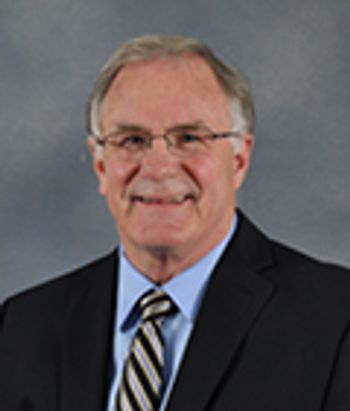
In forensic science, the detection of blood on fabric is a very useful tool. Therefore, it is important that the methods used for detecting blood be as accurate as possible. Michael L. Myrick and Stephen L. Morgan, both professors in the Department of Chemistry and Biochemistry at the University of South Carolina, have been investigating the use of infrared (IR) spectroscopy for this purpose, including comparing the effectiveness of infrared diffuse reflectance versus attenuated total reflectance Fourier-transform IR (ATR FT-IR). They recently spoke to Spectroscopy about their recent studies and the critical questions they have been addressing in how IR spectroscopy is used in forensic science.
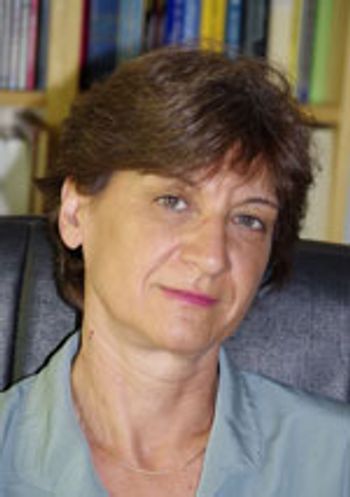
Although laser-induced breakdown spectroscopy (LIBS) potentially can be used for practically any kind of sample, most applications have focused on solid sample analysis. Montserrat Hidalgo, a professor in the Department of Analytical Chemistry and Food Sciences and the University Institute of Materials at the University of Alicante in Alicante, Spain, has been working with various approaches to extend the applicability of LIBS to trace-elemental analysis of liquid samples. She recently spoke to us about this research.
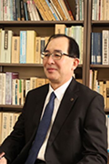
Naoto Nagai, of the Industrial Research Institute of Niigata Prefecture in Japan, has been studying the potential of IR spectroscopy for investigating higher-order structures of polymers. He and his colleagues recently looked at the IR spectra of polyoxymethylene (POM) mold plates and the cause of occasional resin cracks.
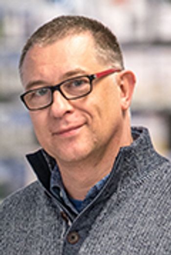
In drug development, quantitative determination of a candidate drug and its metabolites in biofluids is an important step. The standard technique for quantitative metabolite profiling is radiolabeling followed by high performance liquid chromatography (HPLC) with radiodetection, but there are disadvantages to this approach, including cost and time, as well as safety and ethical concerns related to administering radiolabeled compounds to humans.
Surface-enhanced Raman spectroscopy (SERS) with silver nanorod-array substrates has been used in various biological applications, such as detection of proteins in body fluids. Duncan C. Krause, who is a professor in the Department of Microbiology at the University of Georgia, worked with his group to establish a SERS method with those substrates for detecting the pathenogenic mycoplasma that causes bronchitis and pneumonia. We recently spoke with him about this research.
Coherent two-dimensional infrared spectroscopy (2D IR) uses a series of IR femtosecond laser pulses to pump and then probe the response of a system, making it possible to learn much more about the structure and dynamics of molecules than can be seen with one-dimensional IR spectroscopy. The technique’s inventor, Martin T. Zanni of the University of Wisconsin-Madison, discussed 2D IR in a 2013 interview in Spectroscopy (1). Since 2013, Zanni has applied 2D IR spectroscopy to new systems and has started a company, PhaseTech Spectroscopy, Inc., to commercialize the technique.
Proteomics and structural biology require specialized mass spectrometry methods for characterizing protein structures and conformations. Jennifer S. Brodbelt, a professor of chemistry at the University of Texas at Austin, focuses on the development and application of photodissociation mass spectrometry for studying biological molecules such as peptides, proteins, nucleic acids, oligosaccharides, and lipids. She recently spoke with Spectroscopy about her work with this technique. She is the winner of the 2017 ANACHEM Award, which will be presented at the SciX meeting in October 2017. The award is presented annually to an outstanding analytical chemist based on activities in teaching, research, administration, or other activities that have advanced the art and science of the field.
Using Raman and FT-IR spectroscopy for on-line monitoring of manufacturing processes offers advantages such as improved quality control, nondestructive analysis, and reduced costs. Jim Rydzak has more than 20 years of experience leading teams in applying on-line process control, in both the pharmaceutical and consumer goods industries. He recently talked to Spectroscopy about that work, including what they achieved and how they overcame challenges.
Spectroscopy
Multiple-collector inductively coupled plasma–mass spectrometry (MC-ICP-MS) is a powerful technique for measuring isotopic ratios in various areas of research. Michael Wieser, who is an associate professor in the Department of Physics and Astronomy at the University of Calgary, uses MC-ICP-MS to measure isotopic compositions at trace levels in applications ranging from geological studies to protein research. He recently spoke to Spectroscopy about this work.
In biomedical applications of surface-enhanced Raman spectroscopy (SERS), nanoparticles can enhance the Raman signal and provide additional functionality. Duncan Graham of the University of Strathclyde has been pushing the limits of what can be achieved using functionalized nanoparticles and SERS, in applications such as cholera detection, lipid profiling in cancer cells, and assessing the efficacy of anti-cancer drugs, For this and other work he has won the 2017 Charles Mann Award, presented by the Federation of Analytical Chemistry and Spectroscopy Societies (FACSS). He recently spoke to Spectroscopy about this work. This interview is part of a series of interviews with the winners of awards that will be presented at the SciX 2017 conference in October.
Spectroscopy is proud to have created a new award, the Emerging Leader in Atomic Spectroscopy Award. As its name implies, the award recognizes a young scientist, and it is designed to encourage the next generation of atomic spectroscopists. George Chan, the winner of the inaugural Emerging Leader in Atomic Spectroscopy Award, is a project scientist at Lawrence Berkeley National Laboratory (LBNL) in Berkeley, California. One of Chan’s most significant contributions to the field of atomic spectroscopy has been his work on matrix effects and excitation processes in the inductively coupled plasma (ICP).
Optical coherence tomography (OCT) is an emerging technique for medical imaging that uses light to see deep inside tissue. Rohith Reddy, who is a postdoctoral research fellow at the Harvard Medical School and Massachusetts General Hospital in Boston, has worked to develop an OCT device for noninvasive diagnosis of a precancerous condition, Barrett’s esophagus. Reddy is the winner of the FACSS 2016 Innovation Award. He recently spoke to us about these efforts.
Surface-enhanced Raman scattering (SERS) has the capability of enhancing the signal from analytes present in low concentrations, and the detection of drugs present in human and other samples is an important application of this technique. Roy Goodacre is a Professor of Biological Chemistry in the School of Chemistry at the University of Manchester, and he and his group have been developing SERS methods for analyzing drugs in various solutions, including human biofluids, with the ultimate goal of monitoring dosing and drug therapy. He recently spoke to us about this work.

Spectroscopy
Metals and metalloids, while essential to living organisms, can, in high concentrations, be toxic. An understanding of how these metals and metalloids are accumulated and transported within plants and animals is possible with the use of synchrotron X-ray fluorescence (SXRF) microtomography. The technique is used in the imaging of major and trace element distributions within natural materials with high spatial resolution. Stefan Vogt of the Argonne National Laboratory in Lemont, Illinois, has been exploring the use of SXRF to detect metal content in biological and other systems. He recently discussed the various challenges, applications, and advantages associated with this technique.
Advertisement
Advertisement
Trending on Spectroscopy Online
1
Peter R. Griffiths (1942–2026): A Life Devoted to Infrared and Vibrational Spectroscopy
2
How is Raman Spectroscopy Being Used in Life Science Applications?
3
The Current Gaps in Food Science
4
In Memoriam: John Philip Walters (July 4, 1938 - August 18, 2026)
5